


Head office
SECO Sensor Consult GmbH
Am Bleichanger 1
96450 Coburg
Phone: +49 (0)9561 869 141
Fax: +49 (0)9561 869 149
Email: info@seco-sensor.de
Single-particle cryo-electron microscopy (cryo-EM) is one of the most important methods in structural biology today. It makes it possible to visualize the three-dimensional structure of proteins and biological macromolecules with near-atomic resolution. However, despite major advances in detectors, image processing, and sample preparation, one problem remains particularly persistent: the so-called “preferred orientation” of particles.
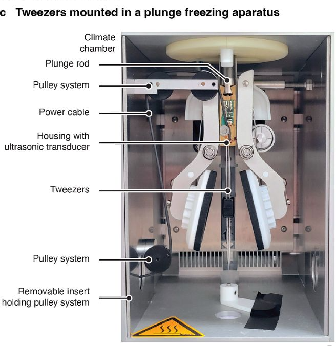
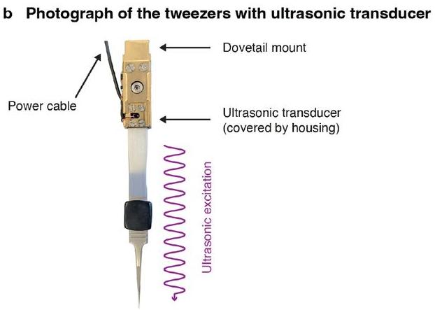
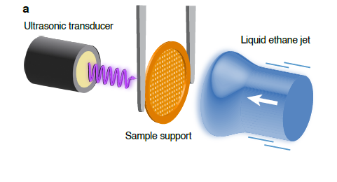
Researchers at the École Polytechnique Fédérale de Lausanne (EPFL) have now developed a simple and practical solution. By integrating our miniaturized SECO ultrasonic transducer SC049 into the tweezers of a standard plunge freezer, they were able to significantly improve the orientation distribution of particles and thus enhance the quality of cryo-EM reconstructions.
To produce a high-resolution 3D reconstruction, cryo-EM requires images of a particle taken from as many different angles as possible. In practice, however, many proteins tend to deposit at the air-water interface of the thin liquid film during sample preparation. As a result, they often adopt similar orientations.
The result: Certain views are overrepresented, while others are barely present or absent altogether. This significantly complicates the reconstruction process, increases the measurement effort required, and, in the worst-case scenario, can prevent the identification of any meaningful structure at all.
To solve this problem, numerous approaches have been developed in recent years, such as tilted data acquisition, special carrier films, or accelerated vitrification processes. However, many of these methods require additional optimization steps or specialized equipment, or come with drawbacks in terms of data quality and throughput.
bioRxiv 2026, Courtesy of: Constantin Richard Krüger, “Plunge freezing tweezers with ultrasonic excitation…”
bioRxiv 2026, Courtesy of: Constantin Richard Krüger, “Plunge freezing tweezers with ultrasonic excitation…”
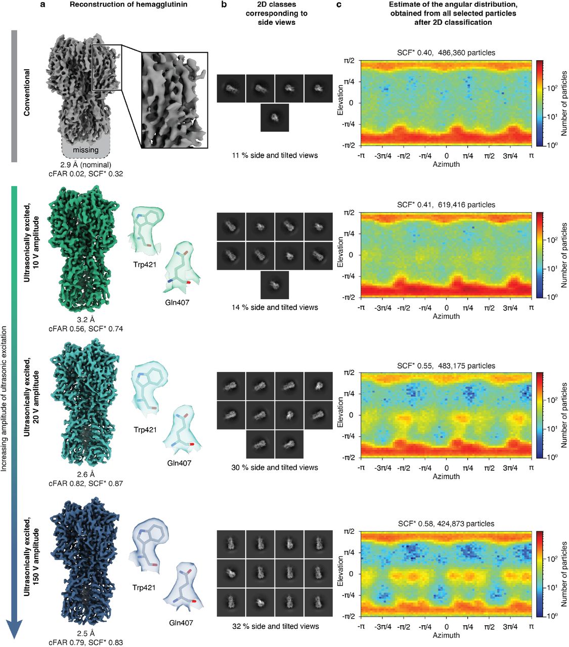
The researchers used the influenza virus protein hemagglutinin as a test subject. It is considered a classic example of strong preferred orientation in the cryo-EM community and is therefore particularly well-suited for evaluating new approaches. With conventional sample preparation, top views of the protein were almost exclusively dominant. Side views accounted for only about 3 percent of the particles, while another 8 percent were slightly tilted views. The resulting 3D reconstruction was incomplete and showed significant artifacts. The achievable resolution was 2.9 angstroms.
bioRxiv 2026, Courtesy of: Constantin Richard Krüger, “Plunge freezing tweezers with ultrasonic excitation…”
An improvement was already evident at an ultrasonic excitation level of 10 volts. More side views became visible, the reconstruction was complete, and it achieved a resolution of 3.2 angstroms.
The best results were achieved at 20 volts. The proportion of side and oblique views rose to about 30 percent, the orientation of the particles became significantly more uniform, and the resolution improved to 2.6 angstroms. With an excitation voltage of 150 volts, the resolution was ultimately increased to 2.5 angstroms. At the same time, it became apparent that higher voltages offer only limited additional benefits in terms of orientation distribution.
In addition to hemagglutinin, the team also studied C-reactive protein and the 50S subunit of bacterial ribosomes. Here, too, ultrasonic stimulation influenced the orientation distribution of the particles. Improvements in reconstruction quality were observed particularly for C-reactive protein. At the same time, the results show that the effect depends on the specific sample and that not every system benefits equally.
A key advantage of the new solution is its practicality. The tweezers modified with the SC049 can be integrated into existing sample preparation systems without fundamentally altering their function. This significantly lowers the barrier to adoption in everyday laboratory work.
bioRxiv 2025, courtesy of Michal Haubner, „Overcoming Preferred Orientation in Cryo-EM With Ultrasonic Excitation During Vitrification“
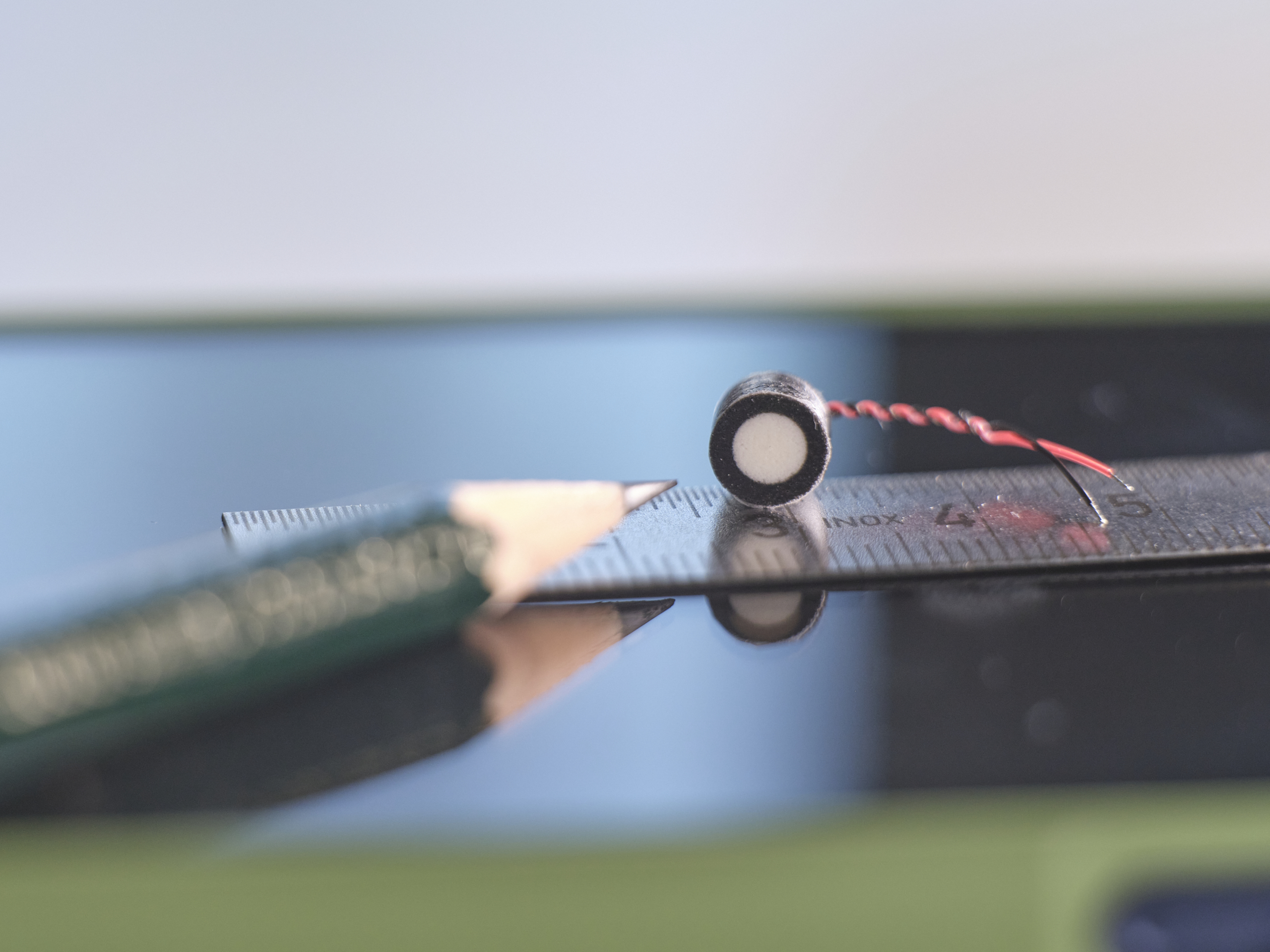
SC049 ultrasonic transducer
Our product manager, Stefan Schneider, would be happy to advise you on our smallest ultrasonic transducer, the SC049. With dimensions of just 6 x 5 millimeters, it is perfectly suited for numerous industrial applications—as well as for complex challenges in laboratories and scientific research.
Upon request, we can customize our SC049 to meet your specific measurement needs, modifying elements such as the housing, connections, or additional features.
Contact us! We look forward to hearing from you.

Stefan Schneider
product manager
SECO Sensor Consult GmbH
Am Bleichanger 1
96450 Coburg
Phone: +49 (0)9561 869 141
Fax: +49 (0)9561 869 149
Email: info@seco-sensor.de

You need to load content from reCAPTCHA to submit the form. Please note that doing so will share data with third-party providers.
More Information